Different imaging and assessment tools across multiple clinics can result in varied prognostic values. Researchers from Japan conducted a retrospective study of harmonized pretreatment volume-based quantitative FDG-PET/CT parameters for prognostic values in breast cancer patients.
The Trending with Impact series highlights Oncotarget publications attracting higher visibility among readers around the world online, in the news, and on social media—beyond normal readership levels. Look for future science news and articles about the latest trending publications here, and at Oncotarget.com.
—
Breast cancer consists of a wide variety of tumor types, symptoms, disease progression courses, and responses to treatments. In the clinic, researchers decide which disease interventions to use by evaluating the patients’ stage of tumor-node-metastasis (TNM), histologic tumor grade, and the levels of hormone receptors and molecular markers that are present.
Standardized uptake value (SUV), metabolic tumor volume (MTV), and tumor lesion glycolysis (TLG) are derived from 18F-fluorodeoxyglucose positron emission tomography/computed tomography (FDG-PET/CT). These variables have also been reported to correlate with clinicopathological prognostic factors and are considered predictive factors of prognosis.
Breast Cancer Prognostic Parameters
“Recently, noninvasive diagnostic tools have been gaining popularity for prediction of tumor behavior, with magnetic resonance spectroscopy (MRS) and diffusion-weighted imaging (DWI) with magnetic resonance imaging (MRI) reported to provide surrogate imaging biomarkers showing correlations with clinicopathological prognostic factors [2, 3].”
In a multi-institutional retrospective study in Japan, researchers—from the Hyogo College of Medicine, Nippon Medical School Hospital, National Cancer Center Hospital, Kinki University Faculty of Medicine, and Gunma Prefectural College of Health Sciences—explain that the factors and algorithms used by different assessment tools across multiple clinics can result in varied standardized uptake values. These inconsistencies have provided an opportunity for the researchers to standardize parameters of prognostic values when imaging breast cancer patients to improve patient outcomes.
“Thus, a harmonization strategy is necessary for comparing semi-quantitative PET parameters among available imaging methods, which is a notably relevant issue for multicenter trials employing different PET systems.”
The Study
Researchers gathered records of 546 patients treated from 2010 to 2016 with stage I–III invasive breast cancer. Of those patients, 344 were estrogen receptor (ER)-positive/human epidermal growth factor receptor two (HER2)-negative, 110 were HER2-positive, and 92 were triple-negative. The patients were treated at four separate institutions using different PET/CT scanner systems. In addition to surgeries, chemotherapy, and radiotherapy, patients were assessed during their follow-up appointments.
“Mammography, ultrasound, CT, bone scanning, and FDG-PET/CT were used for determining disease recurrence, metastasis, and progression during follow-up.”
Researchers in this study retrospectively performed histological and statistical analyses of overall survival and recurrence-free survival in patients of each breast cancer subtype group.
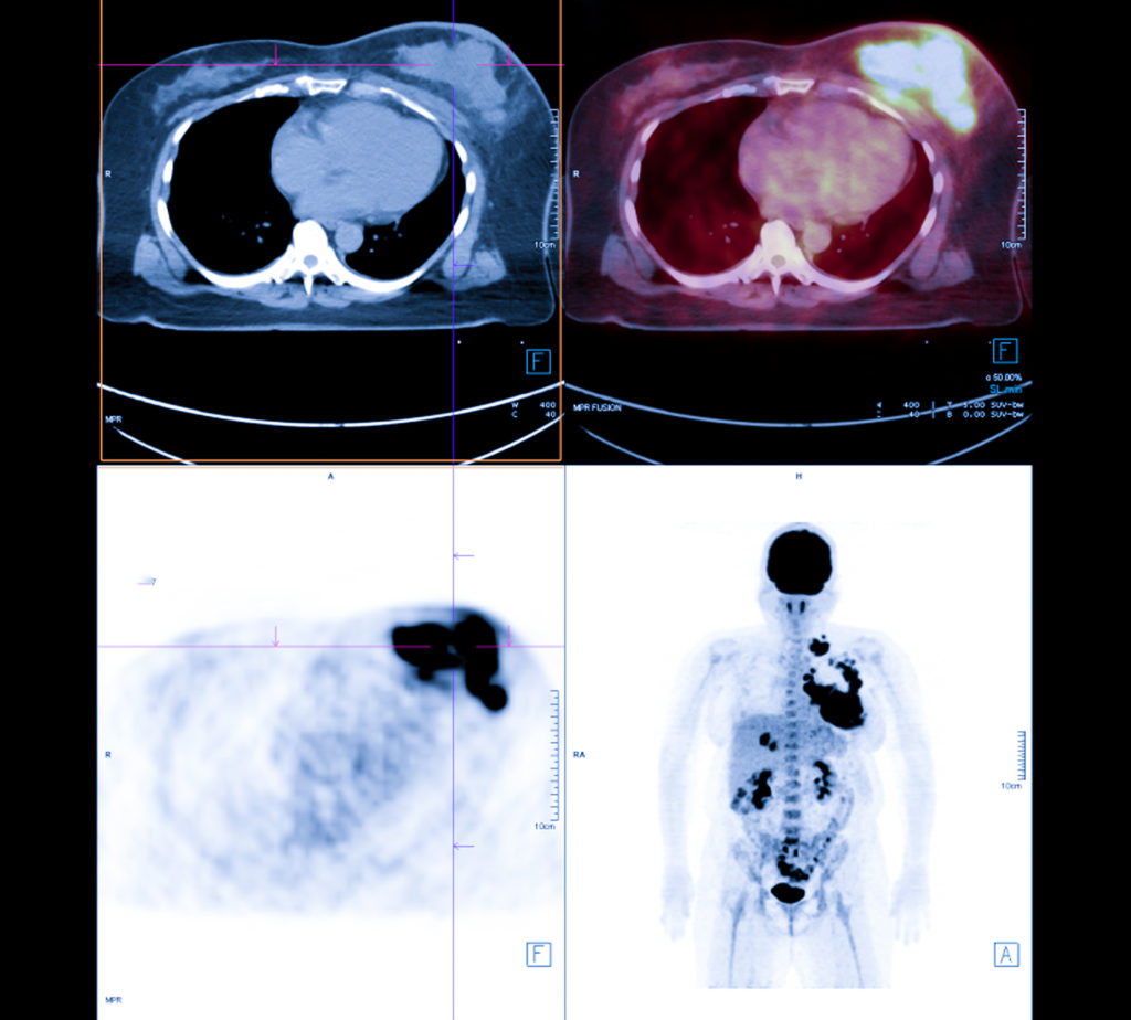
“An experienced reader (12 years of experience with oncologic FDG-PET/CT) who had no knowledge of other imaging results or clinical and histopathologic data retrospectively reviewed all of the FDG-PET/CT images.”
They found that the average maximum standardized uptake values (SUVmax) for HER2-positive and triple-negative tumor patients were higher than in patients with ER-positive/HER2-negative tumors.
“Harmonized primary tumor and nodal maximum SUVmax, metabolic tumor volume (MTV), and TLG indicated in pretreatment FDG-PET/CT results were analyzed.”
Conclusion
Results from this study suggest that harmonized PET classifications with final clinical response assessments demonstrate a better ability to predict disease-free survival compared to non-harmonized PET classification.
“We concluded that harmonized quantitative volume-based values, especially those for the primary tumor and nodal SUVmax and TLG, obtained with FDG-PET/CT can provide useful information regarding prognosis for both recurrence and death in patients with operable invasive breast cancer, including all three main subtypes. The findings presented here are considered useful for improving care of individual patients.”
Click here to read the full retrospective study, published in Oncotarget.
—
Oncotarget is a unique platform designed to house scientific studies in a journal format that is available for anyone to read—without a paywall making access more difficult. This means information that has the potential to benefit our societies from the inside out can be shared with friends, neighbors, colleagues and other researchers, far and wide.
For media inquiries, please contact media@impactjournals.com.